Fold Recognition Example


Ana Rojas Mendoza.
ProteinDesign Group.
Centro Nacional de Biotecnologia (C.N.B.- C.S.I.C.)
Tlf: +34-91-5854570. Fax: +34-91-5854506.
Madrid-Apr-04


3D Structure Prediction: A Fold Recognition Example.

Sequence:
>BX842656_101
MLKNTSSNLDAPVARSCDFAMKKMDLRKAFTLIEPGPVTLVTTSAGGTNNVMTISWTMAV
DFTPKLAITTGPWNFSYKALTKSRECVIAIPTVDLLDKVVGVGTCSGKDTDKFDTFRLTP
IKGKYVEAPLIKECVANIECKVVDIIKKHDIVVLEGVAAYFDTSRKEKRTLHAVGDGTFV
VDGRTLDRKKQMRSKLLGIF


Steps to follow:
0.- 1D data from the target         This is the Predict protein [ OUTPUT]
1.-Is this protein similar to another with known structure?         The results of "BLAST search against PDB.
        There IS NOT a known structure with sequence simmilarity to our query.
2.-What can I get from the annotation?: SwissProtFile.
         In this case, swissprot indicates it is an hypothetical protein containing a Flavin reductase like domain. functions.
3.-Results from some fold recognition steps
4.- How consistent is my fold assignment?          Some of the predicted folds are structuraly related:
FSSP comparison results file
5.a- Fold Recognition on 1EJE_A structure         Based on the secondary structure data, the protein might have 2 domains, one seems to be "all beta" and another the other one "mixed alpha-beta". The active site (FMN binding) of the query protein is located in the same spatial place as the template (1EJE_A) structure.
Alignment structure-sequence : text format
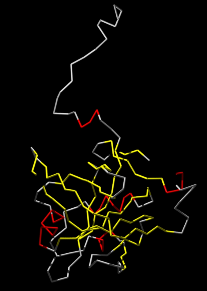
5.b- Mapping the query on 1EJE_A structure

   YELLOW: B-Strands.
   RED: Helices.
   GREY: No secondary structure      elements.
   Note the loops (are missing)

5.c- Fold Recognition on 1EJE_A structure

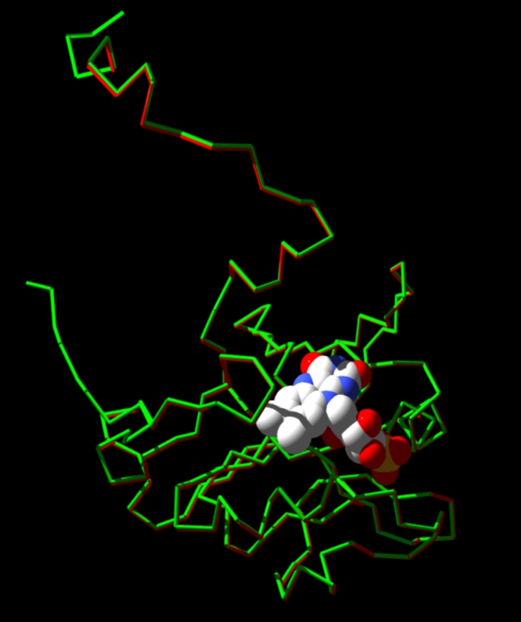
A comparison between the predicted structure and the actual one:

RED: The predicted model.
GREEN: The template.
REST: FMN (flavin) group and Ni heteratms.

And, [HERE],the PDB file of the above comparison structure.